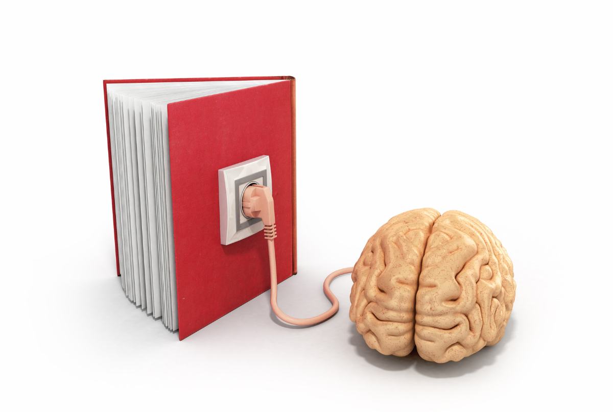
Skilled readers can recognize words at lightning fast speed when they read because the words have been placed in a sort of visual dictionary. When they look at a known word, their brain sees it like a picture, not a group of letters needing to be processed.
That’s the finding from Georgetown University Medical Center (GUMC) studies, which show the brain learns words quickly by tuning neurons to respond to a complete word, not parts of it.
How the brain recognizes words
The existence of brain regions dedicated to reading has been fiercely debated for almost 200 years. Wernicke, Dejerine, and Charcot, among the most important and influential neurologists and neuroscientists of the 19th century, debated whether or not there was a visual center for words in the brain.
We now know there is, and that we are not recognizing words by quickly spelling them out or identifying parts of words, as some researchers have suggested. Instead, neurons in a small brain area remember how the whole word looks – using what could be called a visual dictionary.
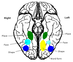
This small area in the brain, called the visual word form area (VWFA), is found in the left side of the visual cortex, opposite from the fusiform face area on the right side, which remembers how faces look. One area is selective for a whole face, allowing us to quickly recognize people, and the other is selective for a whole word, which helps us read quickly.
These findings negate the dual-access theory, which suggests that visual word recognition in skilled readers is not based on visual processing alone but that we access both the phonology and the visual perception of a word (Frost, 1998).
The brain changes in response to nonsense words
A 2015 GUMC study asked 25 adult participants to learn a set of 150 nonsense words. The brain plasticity associated with learning was investigated with functional magnetic resonance imaging (fMRI), both before and after training.
The researchers found that neurons respond differently to real words, such as turf, than to nonsense words, such as turt, showing that a small area of the brain is “holistically tuned” to recognize complete words.
Using a specific fMRI technique know as fMRI-rapid adaptation, the investigators also found that the visual word form area changed as the participants learned the nonsense words. Before training the neurons responded like the training words were nonsense words, but after training the neurons responded to the learned words like they were real words.
Separate brain regions for visual and sound
In a 2016 GUMC study, the researchers tested word recognition in 27 volunteers using fMRI. They were able to see that words that are different but sound the same (like “hare” and “hair”) activate different neurons, akin to accessing different entries in a dictionary’s catalog.
If the sounds of the word had influence in this part of the brain, we would expect to see that they activate the same or similar neurons, but this was not the case; ‘hair’ and ‘hare’ looked just as different as ‘hair’ and ‘soup.’ This suggests that all we use is the visual information of a word and not the sounds.
When we see a word for the first time, it requires some time to read and sound it out, but after perhaps just one presentation of the word, we can recognize it without sounding it out. This occurs because our brain first uses phonology to encode the word and match the sound with the written word. Once we do that and encounter the word a few more times, we no longer need the phonology at first, just the visual input to identify the word.
In addition, the researchers found a distinct region sensitive to sounds, where ‘hair’ and ‘hare’ did look the same. The researchers thus showed that the brain has regions that specialize in doing each of the components of reading: one region is doing the visual piece, and the other is doing the sound piece.
Brain’s VWFA is critical to fluent reading
In the most extensive study of its kind up to date, Brem et al. (2020) confirmed that the VWFA is critical to fluent reading using fMRI data (n = 140 children, 7.9–12.2 years; 55 children with dyslexia, 73 typical readers, 12 intermediate readers). Their study also showed that connectivity of the left occipitotemporal cortex seems a prerequisite for guiding the emerging specialization in the VWFA.
The VWFA is insensitive to surface visual characteristics, such as case, font, size, and length, meaning it takes only the relevant information. For example, this area recognizes TABLE, table, tAbLe, and table as the same word. It is also insensitive to orthographic regularity (activating equally for regular and irregular words). These characteristics allow the VWFA to support easy, fast, effortless, and automatic word recognition in literate individuals.
Research by Pedago et al. (2011) supports that the VWFA plays a crucial role in the discrimination of letter orientation, which links to the well-known fact that a deficit in discriminating between a b and a d is a common symptom of dyslexia.
Given that the most robust underactivation in children with dyslexia occurs in the left occipitotemporal cortex (Brem et al., 2020; Martin et al., 2016), this brain region should be a prime target for supportive interventions.
Edublox offers live online tutoring to students with dyslexia that targets both the visual and sound pieces in the brain. Our students are in the United States, Canada, Australia, and elsewhere. Book a free consultation to discuss your child’s learning needs.
.
.

.